





 By
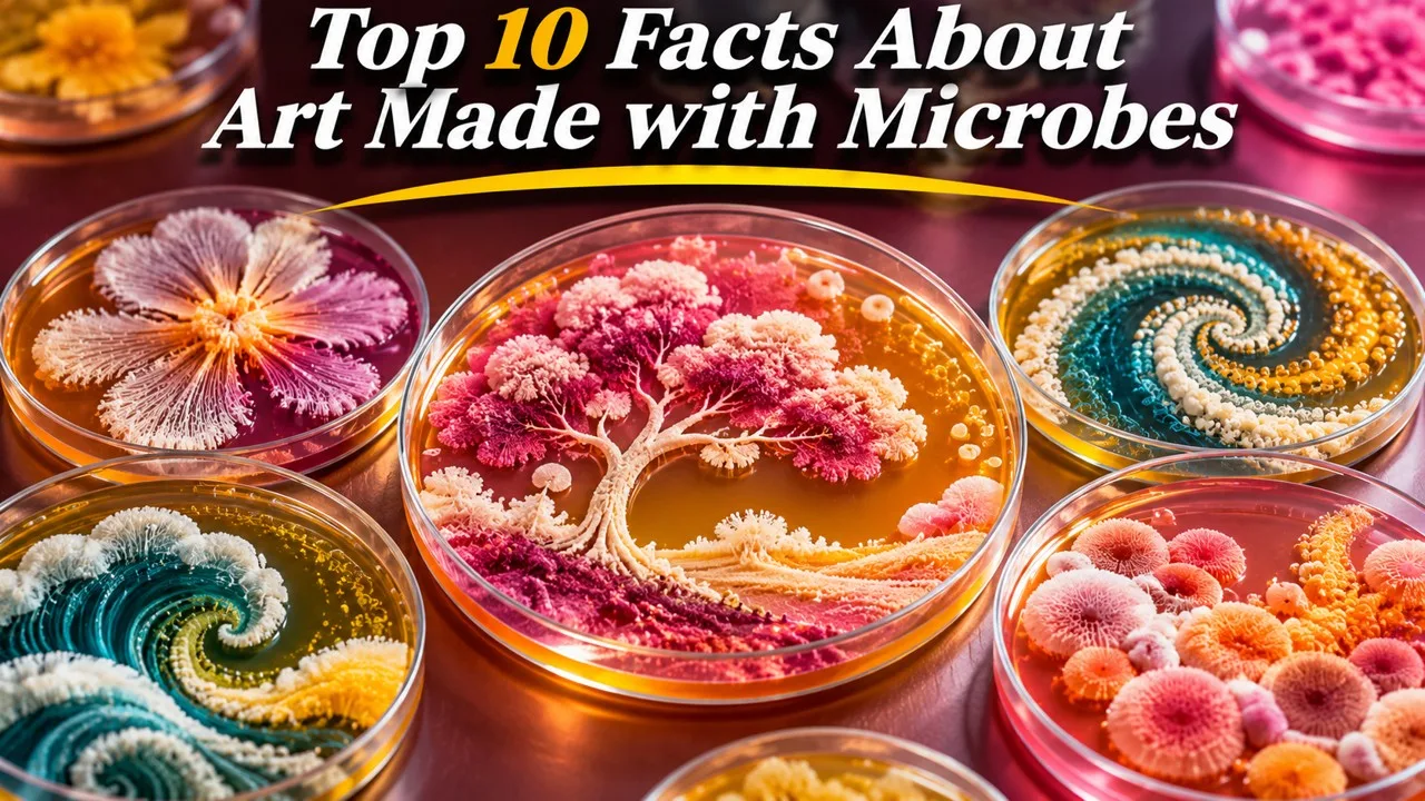
Agar, pigmented colonies and a yearly contest have made microbes into a medium; the works expire, and they are not a reason to culture unknown bacteria at home. Microbial art is a real practice with a history, a set of materials, and a public contest, not a secret underworld of living paint [4][5]. It is also a laboratory activity. Some of the most photogenic pigmented species include organisms that can be opportunistic pathogens, and this article will not provide a method, a recipe, or a shopping list for growing them [3]. The ten facts below describe the field as it is actually practiced in trained labs and in documented exhibitions.
The pictures that matter, for a public article, are photographs of plates that have already been made under supervision, not a prompt to start a kitchen culture.
Key Facts
Microbial art uses living colonies as color and line. The work is grown, not only applied, and it has a lifespan.
Ten Facts at a Glance
| Rank | Fact | The careful version |
|---|---|---|
| 1 | The medium is usually agar in a dish | Nutrient jelly, a sterile plate, and a controlled inoculation, not a household saucer |
| 2 | Colonies can be "painted" by inoculation | A loop or swab places cells in a design; they then grow into the visible work |
| 3 | Color often comes from the organisms themselves | Pigmented bacteria and fungi supply reds, yellows, and other hues |
| 4 | There are real risks | Some pretty species can infect a compromised host; trained biosafety is the point, not a how-to |
| 5 | The work expires | Colonies dry, change, overgrow, and are often destroyed after being photographed |
| 6 | Alexander Fleming made pictures with bacteria | Decades before a formal contest, he used pigmented colonies as a drawing medium |
| 7 | An annual agar-art contest exists | The American Society for Microbiology has run a public contest since 2015 |
| 8 | "Good" microbes are still not toys | Educational art can show useful or harmless species without erasing caution |
| 9 | The field sits between lab and gallery | Bioart programs and science museums have given the practice a public room |
| 10 | The possibilities are not literally endless | New pigments and safe strains appear, within the limits of ethics and biosafety |
1. Jelly, a Dish, and a Sterile Habit
Most microbial pictures are made on agar, a seaweed-derived jelly poured into a Petri dish and loaded with nutrients [1]. The plate is a tiny garden with a lid. In a proper laboratory it is poured and handled so that only the intended organisms grow, which is why a kitchen counter is not a studio for this medium [1]. The "necessary materials" are therefore ordinary microbiology tools, not a secret palette, and they include the unglamorous ones: a way to sterilize, a way to dispose, and a person who knows what they have plated.
The art begins after that housekeeping. Without it, the dish is only a risk, and a pretty risk is still a risk. That is the first fact, even if it is the least glamorous one on the list.
2. Drawing with a Living Line
The artist does not, in the usual case, smear a finished pigment and walk away. They inoculate: they place living cells in a pattern, often with a loop, a swab, or a stamp, and then they wait while colonies grow into visible points, lines, and washes [2]. Timing and temperature are part of the composition, because a plate left too long will blur as colonies spread and overtake one another [2]. The closest analogy is a garden that has to germinate in the shape of a drawing.
This is why photographs, taken at the right hour, are often the work that the public sees. The plate itself is a performance with a peak.
Related: Top 10 Turning Points in the History of Biology
3. Color That Grows
Many of the most striking plates use organisms that make their own color: reds, yellows, oranges, and occasional blues or greens, depending on the species and the medium [3]. The color is a metabolite or a structural effect, not a tube of paint. That fact is the charm of the medium, and also the reason this article will not name a casual home recipe. Some well-known pigmented bacteria have caused infections in hospitals and in other vulnerable settings [3]. A public description can say that pigment is biological. It should not become a collecting guide.
Fungi, yeasts, and mixed safe teaching strains are also used, especially when the aim is a classroom plate rather than a shock of red.
4. The Risk Is Not a Selling Point
Microbial art is sometimes advertised as if the danger were part of the glamour. The honest version is duller and more serious. Anyone who grows environmental or clinical microbes can, in principle, grow something they did not intend, and some pigmented species used historically in displays are not harmless [4]. Teaching labs use approved strains, sealed plates, and disposal by autoclave or an equivalent method. This article will not explain how to culture an unknown sample from a shoe, a sink, or a wound, and it will not treat a pretty colony as proof that an organism is safe.
The risk is why the practice belongs in a lab with rules, not in a viral "try this at home" card.
5. An Exhibition with an End Date
A living plate does not stay in the state the artist wanted. Colonies age, dry, change color, merge, and sometimes sporulate; mold can arrive as an uninvited collaborator [5]. Many works are photographed at their peak and then destroyed, which is both a biosafety step and an aesthetic fact: the art has an expiration date [5]. Archives of microbial art are therefore often archives of images, plus a few preserved or dehydrated curiosities.
Related: Top 10 Most Bizarre Phobias: Strange Fears That Exist
Impermanence is not a failure of the medium. It is one of its subjects, whether or not the artist intended a lesson about time.
6. Fleming's Pictures, before a Contest
Alexander Fleming, already a microbiologist and later famous for penicillin, made drawings with pigmented bacteria in the 1920s and 1930s, including little scenes and figures grown as colonies on paper or on media [6]. He is often called a father of microbial art, which is fair as a genealogy and too grand if it implies a school with students [6]. The pictures were a form of play by a scientist who lived among plates. They were not a gallery movement, and they do not need to be recast as outsider masterpieces.
They do show that the idea is older than the hashtag, and older than the annual contest that now travels with the subject. A scientist at play, among plates he already understood, is a different figure from a hobbyist copying a pattern without that background.
7. A Yearly Contest, with Rules
Since 2015, the American Society for Microbiology has run an Agar Art contest, open to entries made with living microbes and judged in public categories [7]. The contest has done more than any single laboratory to give the practice a yearly calendar, a set of photographs, and a reminder that the work should be done with documented organisms and with safety in mind [7]. This article will not reproduce contest entries, which are other people's pictures, and it will not treat a society logo as part of a featured image.
A contest does not make a medium serious by itself. It does make the medium legible to people who will never enter a teaching lab, and it gives editors a yearly occasion to explain that a plate is both a picture and a culture that has to be treated as both at once.
Related: The Evolution of Stranger Things: Seasons, Themes, and Characters
8. Useful Microbes, Still Handled with Care
Popular writing likes to say that bacteria "are better than they seem," meaning that most species are not villains and that some are essential to soil, food, and health [8]. That is a fair correction to germ panic, and it is a good subject for a teaching plate. It is not a reason to drop caution. An educational work can use yeast, approved laboratory strains, or clearly identified harmless organisms, and still be beautiful [8]. The art is not a moral rehabilitation of every microbe.
The better lesson is proportion: a world of necessary organisms, and a small number that a plate is not the place to advertise.
9. When a Plate Leaves the Lab
Over the last few decades, bioart programs, science museums, and some galleries have shown living or once-living microbial work, often with a conservator's problem attached: how to exhibit a thing that should not be opened [9]. Artists and microbiologists have collaborated, and the results range from pedagogical plates to more ambitious installations about antibiotic resistance, food, or the microbiome [9]. "Merging science and art" is a slogan. The interesting part is practical. Who keeps the lid closed, who signs the safety form, and what is left when the show comes down.
The public can see the work as a photograph, a sealed dish, or a time-lapse, and still understand the point.
Related: Chernobyl Miniseries: Stories of Tragedy of a Nuclear Catastrophe, Heroism and Sacrifice
10. New Pictures, with Limits
There is always another pigment, another safe strain, another way to stencil a plate, and even work that uses yeast to grow a pattern over days [10]. "Endless possibilities" is the language of a press release. The real limits are the interesting ones: ethics, biosafety, the short life of a colony, and the fact that a living medium will not do everything a brush will do [10]. Those limits are also why the good plates are striking. They had to be grown, not only wished for.
The field will continue. It will continue best if it stays a laboratory art with a public face, not a home experiment with a pretty risk, and if the public story keeps the lid, the disposal, and the trained hand in the same frame as the color.
Conclusion
Microbial art has materials, a method of inoculation, a short life, a famous early practitioner, and a yearly contest that brought it to a wide audience. It also has a safety rule that a list should not wink away. Some of the most colorful organisms are not casual pets, and an unknown culture is not a paintbox.
The facts worth keeping are therefore double. Colonies can be a medium, and they can be photographed, taught, and exhibited with care. They are not a reason to grow a mystery sample at home, and they are not improved by a claim that the possibilities have no end. The dish already has enough in it: color, time, and a lid that should stay closed. That is a complete medium, and it does not become more complete by being taken home.
- ^ [1] Standard descriptions of agar media and Petri-dish culture, as used in teaching laboratories. Britannica: Agar
- ^ [2] Educational and ASM materials on agar art as a pattern of inoculation that grows into a visible design. American Society for Microbiology
- ^ [3] Overviews of naturally pigmented bacteria and fungi, including notes that some pigmented species can be opportunistic pathogens. Britannica: Bacteria
- ^ [4] Laboratory biosafety principles for handling cultures, without methods for isolating unknown or hazardous organisms. CDC biosafety resources
- ^ [5] Practitioners' and contest accounts of photographing plates at peak growth and then destroying them. ASM Agar Art
- ^ [6] Historical notes on Alexander Fleming's bacterial drawings in the 1920s and 1930s. Britannica: Alexander Fleming
- ^ [7] American Society for Microbiology Agar Art contest, founded in 2015. ASM Agar Art Contest
- ^ [8] Educational summaries of the many non-pathogenic and useful roles of microbes, paired with ordinary laboratory caution. Britannica: Microorganism
- ^ [9] Surveys of bioart and museum practice involving living microbial works. Britannica: Bio art, where available, and museum education pages
- ^ [10] Discussions of new teaching strains and the ethical and safety limits of living media. American Society for Microbiology

















Discussion
Log in with